2021.4.15~18:The 121st Scientific Meeting of the Japan Society of Medical Physics
The 121st Scientific Meeting of the Japan Society of Medical Physics (JSMP) has been held in PACIFICO Yokohama, Yokohama, Japan. Ms. Umeda (M2) and Ms. Mouri (M2) have attended it for oral presentation.
■Date: April 15 – 18, 2021
■Venue: PACIFICO Yokohama, Yokohama, Japan
■Conference name: The 121st Scientific Meeting of the Japan Society of Medical Physics
Mariko Umeda(M2)
We are pleased to report that our second-year master students Umeda and Mouri gave oral presentations at the 121st Annual Meeting of the Japanese Society of Medical Physics. Due to the spread of the new coronavirus, it was not possible for the students to go to the venue to give their presentations, so they had to submit their slides in advance.
This year’s conference will be held under the theme of “Milestones and Beyond,” in conjunction with the 80th Annual Meeting of the Japan Radiological Society (JRS), the 77th Annual Meeting of the Japanese Society of Radiological Technology (JSRT), and the International Technical Exhibition of Medical Imaging (ITEM2021) of the Japan Industries Association of Radiological Systems (JIRA). The exhibition was held jointly with the 77th Annual Meeting of the Japan Society of Radiological Technology (JSRT) and the International Technical Exhibition of Medical Imaging (ITEM2021) of the Japan Industries Association of Radiological Systems (JIRA). In addition to the local event held at Pacifico Yokohama from April 15 to 18, it was also held on the web from April 28 to June 3.
I made a presentation on “Development of prognostic prediction method with the novel radiomic feature based on graph theory” at JSMP. The presentation was entitled “Development of prognostic prediction method with the novel radiomic feature based on graph theory. Radiomics is one of the prognostic tools for predicting patient outcomes by extracting features from medical images. Currently, many reports use indices such as tumor size, distribution of pixel values on the image, and inhomogeneity as features. As a new approach, we have developed a feature set based on a mathematical concept called graph theory. In this analysis, we compared the prognostic accuracy of graph-theoretic features with that of features obtained by conventional methods, and evaluated whether graph-theoretic features are useful in predicting patient prognosis. The results showed that the prognostic accuracy was improved compared to the conventional method, but we felt that further improvement in prognostic accuracy is necessary to consider clinical implementation and that there are many points that need to be improved.
There were other Radiomics-related presentations at the conference, including reports on the application of Radiomics not only for predicting patient prognosis but also for evaluating dose distribution and predicting pulmonary inflammation after radiotherapy. All of these reports used different approaches and methods from my own research, and I found their ideas and perspectives very helpful.
Finally, I would like to apply the experience and knowledge I gained from this presentation to my future research and conference presentations. I would like to take this opportunity to thank my supervisors and everyone who gave me the opportunity to present my paper.
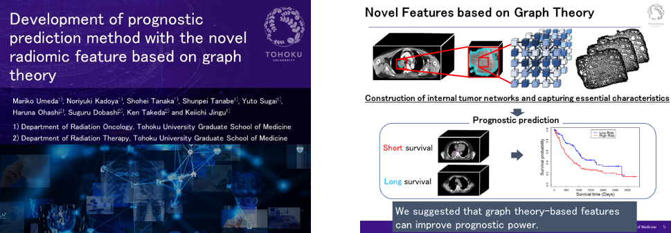
Ms.Umeda’s presentation (Applying Graph Theory to Radiomics)
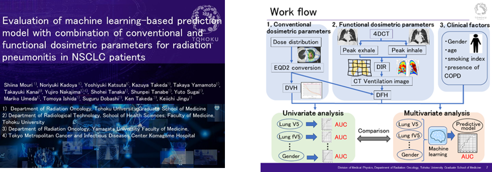
Ms.Mouri’s presentation (Research Workflow)